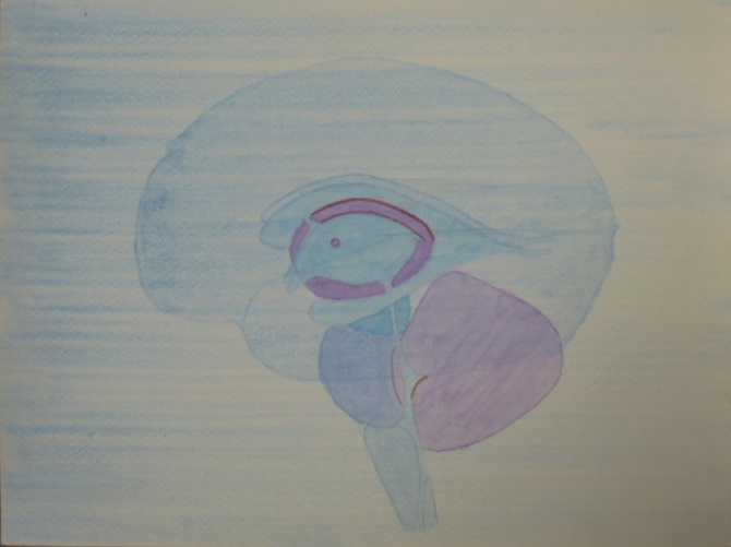
In these pictures and article, I show the energies and dynamics of the cranial sea and brain. These images are wonderful for understanding this part of us that connects us with the higher self and subtle realms.
Having the pictures helps us to visualize this area for self-healing. 
The cranial fluid acts as a medium to receive higher energies.

The anatomy and picture story of the Cranial Sea

The brain is 90% water and the fluid in the brain is 99% water. I call this fluid the cranial sea.
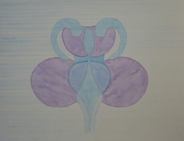
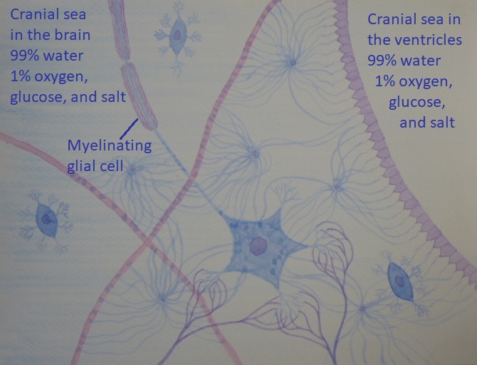
The main body of the brain is full of glial cells (brain cells) these are not close together but are joined by tentacles and a bit similar to seaweed and sea anemones.
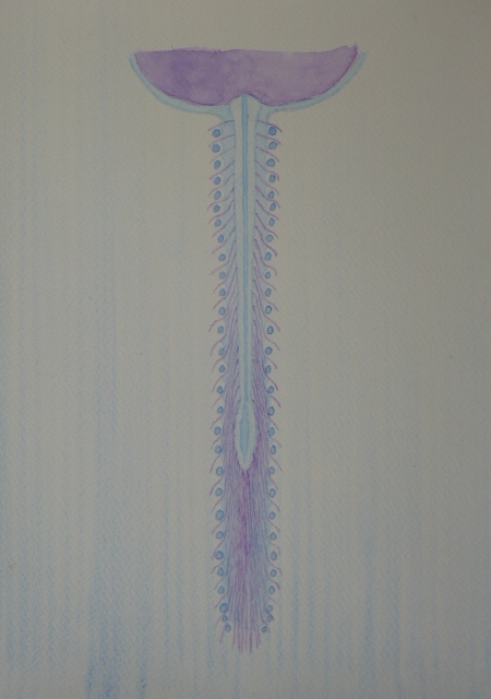
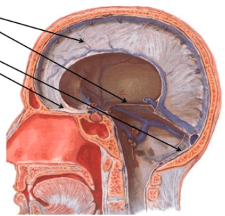
Then there are the ventricles, these are like caverns in this sea without any sea anemones or seaweed in them. The ventricles are just full of cranial sea and nothing else.
The cranial fluid in both the body of the brain and in the ventricles is 99% water and then 1% oxygen, glucose, and salt.

How the cranial sea is made
The three filters that make the cranial sea
This first filter is through the endothelial cells

The cranial sea is 99% water and one of the fundamental principles of water is that it will flow through any membrane and any cell either directly through channels or by osmosis.
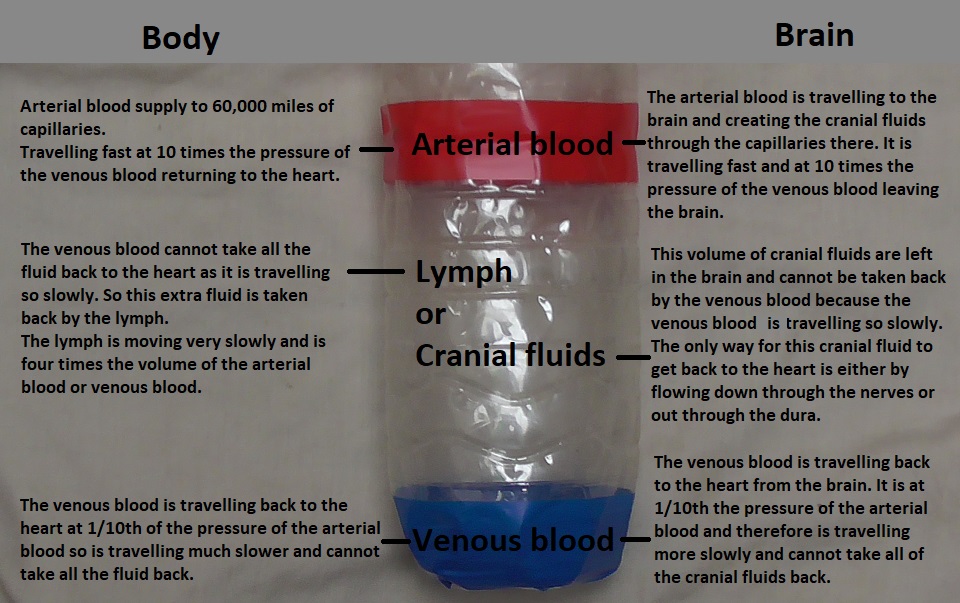
In the brain, the cranial sea is filtered from the blood.
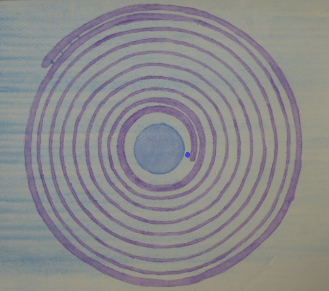
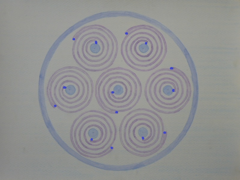
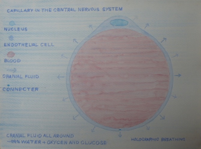
It is filtered through a membrane of cells around the capillaries. These cells have tight junctions. Tight junctions are so close that only 100% pure water can flow through them. As the blood pressure is higher inside the capillaries water will also pass through the body of the cells by osmosis.
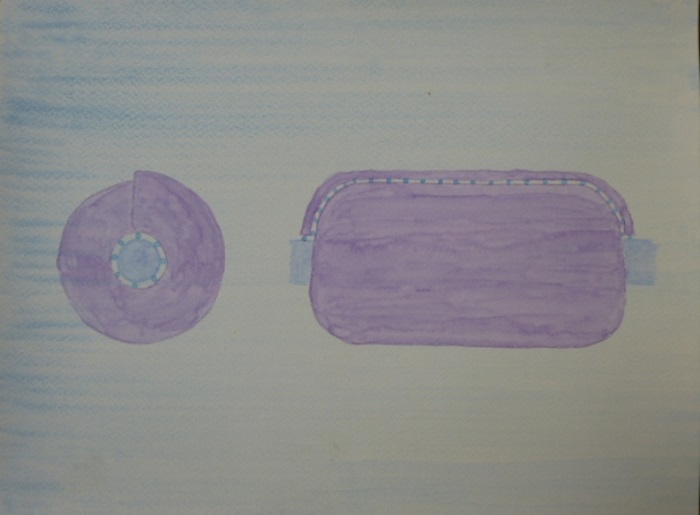
The cells around the capillaries are called endothelial cells. Below is a cross-section of the capillary. It is so small that one endothelial cell can wrap all the way around it. Where it joins together at the bottom, there is a little blue dot, this is the tight junction. Only pure water will flow through this junction. The rest of the water flows directly out through the cell walls.

Endothelial cells are also intelligent they choose which constituents are needed for the cranial sea. These are oxygen, glucose, and salt. The endothelial cell gives the oxygen, glucose, and salt safe passage through its body and out into the cranial sea. The endothelial cell will not let anything else from the blood move through into the cranial sea so it produces a very fine filter.
This means that the brain and glial cells end up in a very pristine fluid environment of 99% water and 1% oxygen, glucose, and salt.
The oxygen and glucose are so the cells can breathe and have food. The salt produces the electrical charges that move along the nerve pathways.

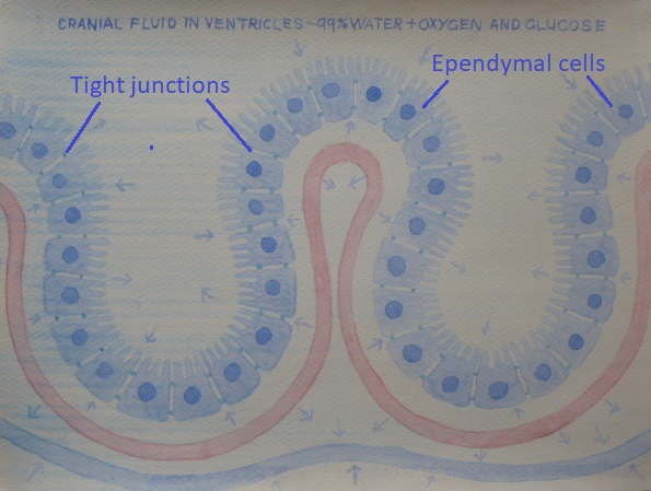
This 2nd filter is through the ependymal cells

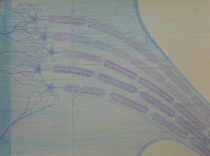
the cranial sea in the ventricles is created from the choroid plexus. The choroid plexus are capillaries that are encased in ependymal cells. Fluid comes out of the arterial capillaries into the space inside the choroid plexus.

The ependymal cells have the same characteristics as the endothelial cells. These cells have tight junctions so only pure water can flow through the junction. Then as the pressure is greater inside the choroid plexus the water will flow out directly through the bodies of the ependymal cells. This will fill the ventricles with pure water.
Then in the same way as in the endothelial cells, the ependymal cells allow safe passage for oxygen, glucose, and salt to travel through their bodies and will not allow anything else to travel through.
So you end up with exactly the same pristine environment created from two different areas, 99% water and 1% oxygen, glucose, and salt. 
Below I show the ependymal cells lining the capillaries.

Here are some pictures of the ventricles this is a side view.

Here they are integrated into the brainstem. They are wrapped around and inside the thalamus and brainstem. The red lines are the choroid plexus.

Here is a front view the 4th ventricle is flat from the sides but from the front makes a diamond.

Here they are including the 5th ventricle this isn't normally shown. Notice how the stem completes them and gives them an earth. The Left-hand one is face on and the right-hand one is from the side. For me the left-hand one is more female and the right-hand one more male. They always look a bit like Mr and Mrs ET to me.

Here they are front and sides integrated into the spine and brainstem.

Here they are on their own front view relating to the kidneys, adrenals, bladder, and urethra, from the blue energy circuit

This 3rd filter is through the myelinating glial cells

The myelinating glial cells are shown in the picture at the top of the page of the cranial sea they line the nerve fibre (axon). Also below is a cross-section.
The myelinating glial cells are filtering cranial fluid for their internal environment. They make spirals with their bodies and the gaps between the spirals they fill with the cranial sea.

All of the connections in these cells have tight junctions so only pure water will flow in through those and only pure water will flow through their bodies.
These cells are also intelligent in the same way as endothelial cells and ependymal cells.
They allow safe passage for oxygen, glucose, and salt. The oxygen and glucose are to feed themselves and the salt is to help fire the electrical charge through the nerve fibre (axon) that they are surrounding.

In the nerves that are travelling out from the brainstem and spinal cord, the myelinating glial cells join together to maintain their internal environment of cranial sea around the axons. They also, make capillaries for the cranial sea to flow through the length of the nerve.

Here is a cross-section of a myelinating glial cell. The little blue dots at the beginning and end of the coil are tight junctions.


Here is a picture of the myelinated nerves leaving the central nervous system

Here is a picture of the nerve fibres and myelinating glial cells travelling out through a nerve. Notice how there's nothing at all to stop the cranial sea flowing out through the nerves they are just open vessels. This was the first picture I painted. Probably by this stage the myelinating glial cells have joined together so there is no gaps with the axon showing. This protects the axon in the nerve so that it is living in an environment of cranial sea. Also as they join together they make capillaries for the cranial sea to travel down the nerves.

Here is a cross-section of the myelinating glial cells travelling down the nerve. The little blue dots are tight connections so only pure water can flow into the cells. This protects its environment of the cranial sea as it travels down through the nerve. The myelinating glial cells also joined together so that the axon is only in contact with cranial sea. These also become capillaries for the cranial sea to travel down through the nerves. In the nerve the blood supply creates normal body interstitial fluid rather than cranial sea. So the myelinating glial cell protects itself and the axon in its own environment of cranial sea. The same as inside the central nervous system.

Here are the myelinating glial cells and axons travelling down the nerves. Where these connect together they create thousands of tentacles that join back-and-forth from both sides. These create a massive surface area for the cranial sea to flow from one myelinating glial cell into another. For me this is very similar to the valves in the lymph system. These capillaries are perfect for capillary action to draw the cranial sea along them. As the cranial fluid is nearly pure water this makes it the heaviest fluid in the body and as most of the nerves are travelling downwards this helps this action.
The myelinating glial cells are about a hundred times as long as they are wide.  As they are so much longer this also helps with the capillary action. 

The image below shows a myelinating glial cell coming to an axon. The blue dot is the cell's nucleus. The cell starts wrapping around the axon and as it travels it filters cranial sea into itself. So it creates a spiral of itself and a spiral of cranial sea. It will keep wrapping around the axon growing in length sideways. Fully grown these can be up to a hundred spirals in the middle and at the ends they will just be one spiral. These cells become so long that it's not possible to show it in a picture fully grown they are about a hundred times as long as they are wide and the same width all the way along.

In the picture below you can see the unravelled sheet of cranial sea that would be between the different layers of the myelinating glial cell. The very bottom centre of the top semicircle is where it is on the surface of the axon in the centre of the myelinating glial cell. Then as it wraps around it gets longer and longer as it spirals out and along the cell. So the top length of the semicircle will be the outside spiral of the cranial sea in the myelinating glial cell.

In the picture below it shows how the sheet of cranial sea spirals out along the axon itself. The areas of blue along the axon are where the cranial sea is on the surface of the axon. The spiralling line on the axon is where physical sheet of the myelinating glial cell is attaching to the axon. So this gives a spiral getting finer as it travels out to either end of the cell. My guess is that this spiral is in line with sacred geometry. Where these myelinating glial cells join together in the nerves and makes capillaries the cranial sea spirals in and out of them as it travels down through capillaries. My internal image is that the electrical charge from the axons spirals out through these fine layers of water and makes light. The four purple drops are the connections of the physical membrane of the myelinating glial cell connecting to the axon. At the sides these are close together and as they move inwards they will spread out more and more with the opening spiral.

Here is a picture of the cranial sea spiralling into and out of the central reservoir. In reality, the thickness of the cranial sea spirals will be thin at the sides and then gradually get wider and wider until they merge into the cranial sea in the middle. Also the outer ones will extend out vertically and then around and join together as the outside horizontal sheet. The 2nd ones will expand out into the 2nd sheet and so forth. 
The axon travels through the middle

For me when they are just shown as the cranial sea they are much more meditative.

Here it is from the outside. The blue dots are tight junctions only letting in pure water. Also, only pure water will flow in and out via osmosis through the outer layer of the cell. And the cell will choose oxygen, glucose and salt to feed itself and the axon. The salt will be used probably in the central reservoir to create the electrical impulse. As I said before these cells are much longer in reality up to a hundred times as long as they are wide. Also in the middle, they can be up to a hundred coils.

I hope you've enjoyed my article and that it may help with your meditation and visualisation.
I will carry on to include the dura in the next article.

About the dura, and the detoxification of the brain and the cranial fluid

Understanding how this all works has been a truly significant part of my journey of healing from Lyme disease. As the spirochetes (the spiral bacteria) of Lyme disease bore into the dura as a place to hide from the immune system and to live. This inflames the dura and also makes it much tougher losing its breathability. I experienced a lot of the deep tiredness from Lyme disease as coming from the dura. When I started using Holographic Breathing to allow the dura to breathe and to detoxify my brain, the tiredness and infection started to leave.
I think many aspects of tiredness diseases are held in the brain, cranial fluid, and dura. In Holographic Breathing, I show how to properly ventilate and detoxify the brain using both the dura and nerves. 
I highly recommend these webinars to anybody but especially to people with Lyme or tiredness issues.

In this picture, I show the central principle of the drainage of cranial fluids. This drainage is primary for our health and detoxification of the brain.

The only way for cranial fluids to leave the brain or spine other than by the venous blood is to leave through the dura and the nerves which are the outer face of the central nervous system. This means that the central body of cranial fluid shown in the picture has to leave via the dura and nerves.
The cranial fluids are 99% water and the fundamental principle of water in our body is that it can flow through any cell or membrane anywhere in the body this includes the dura. They're now also finding that there are lymph ducts in the dura that offload the cranial fluid.